This document summarizes plants’ abilities to sense their environment, and then process and transmit that information, using electrical signals, to different parts of their body and to other plants. We start with intriguing and little known similarities between plants and animals.
Scientists believe that the common evolutionary ancestor of plants and animals could swim, and had “eyes” (eyespots) that let them stay away from shadows and paddle towards light [1–3]. A relic of this proto-plant (and proto-animal), Mesostigma viride, can be found today swimming on microscope slides in biology classes, swinging its whip-like “arms” (flagella) to follow the flashlight beams of school children (Figure 1).
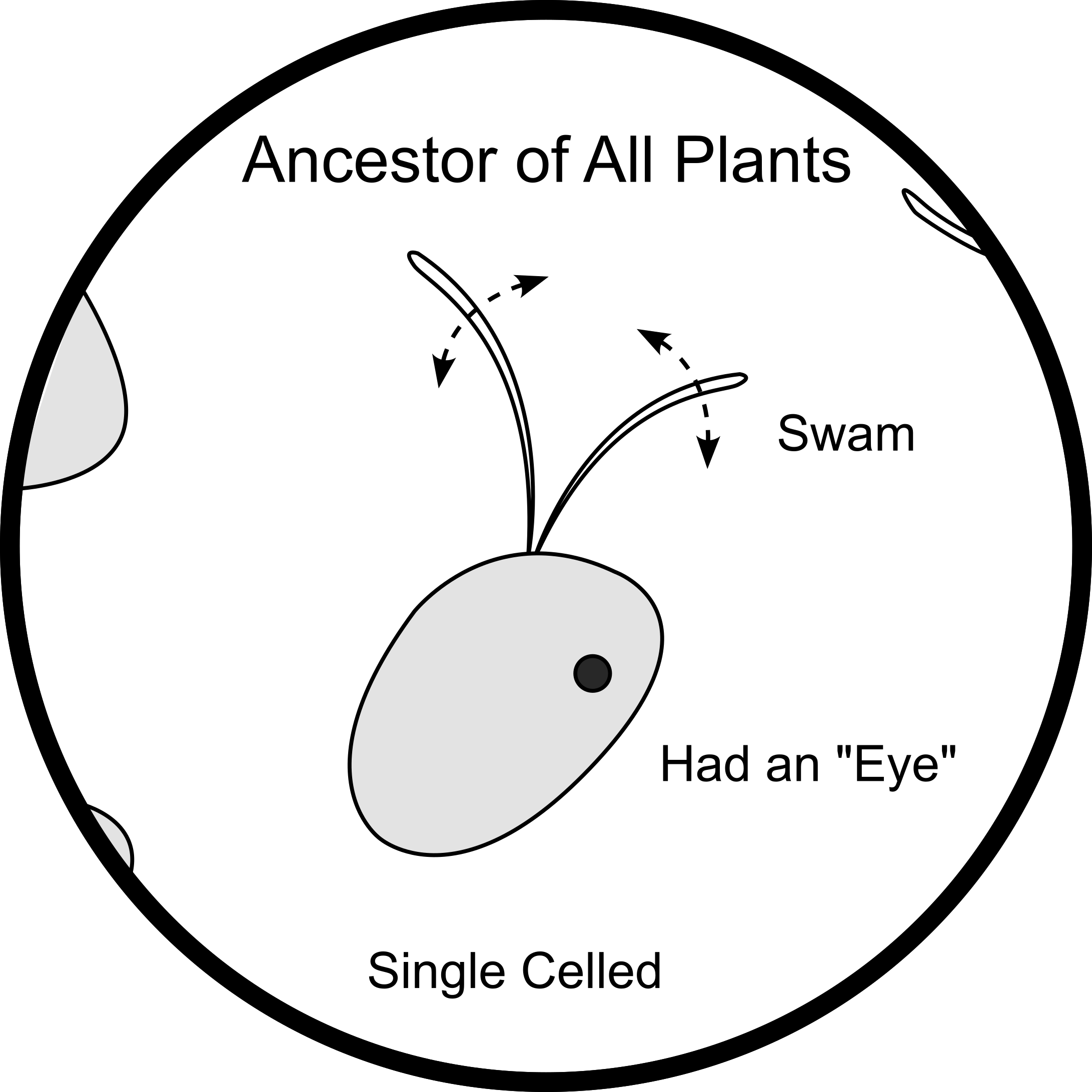
Although modern plants have evolved to move in less obvious ways, they still use actin and myosin (what animal muscles are made of) to transport molecules within their cells [4], and they use these same “muscle proteins” to power the motors in their sperm cell tails, just like animal sperm [5]. So, while modern plants don’t move externally as much as animals do, they have a lot of the same internal moving machinery [6].
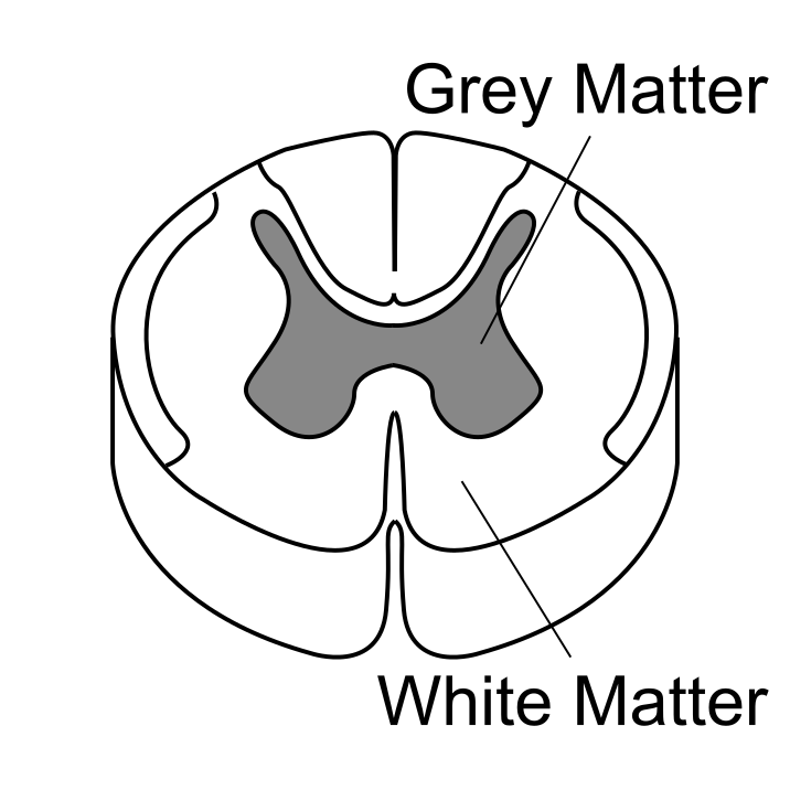
The evolutionary ancestors of plants and animals, mentioned above, were active swimmers that could follow light, but they did not have a nervous system to control their movement. This is because they were single celled organisms, with only a tiny distance (the ~10 micron diameter of a single cell) over which information needed to be conveyed. Over such small distances passive diffusion of chemical signals was sufficient to transfer information from one end of the cell to the other. Only after plant and animal evolution diverged, and each branch evolved into larger, multicellular organisms, did plants and animals each evolve their own long distance signal transmission systems, which ended up being surprisingly similar in many ways. Both plants and animals evolved to encode and transmit information through a cable of specialized cells which traverses the length of their body. In animals, this “cable” is called a nerve, which is made up of individual neuron cells (Figure 2, Left). In plants, the cable is called phloem, which is made up of individual sieve element cells (Figure 2, Right).
Animal
Plant
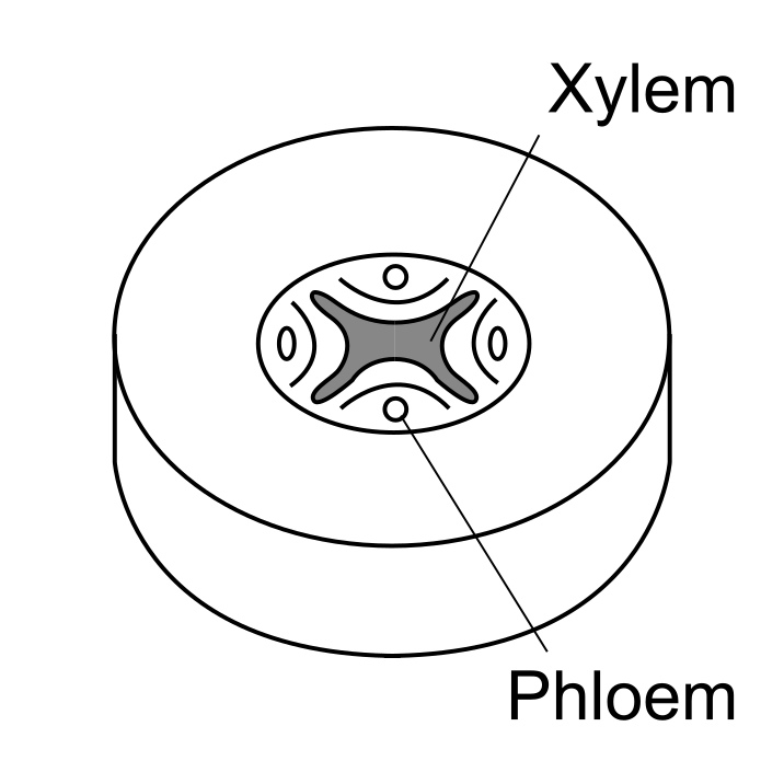
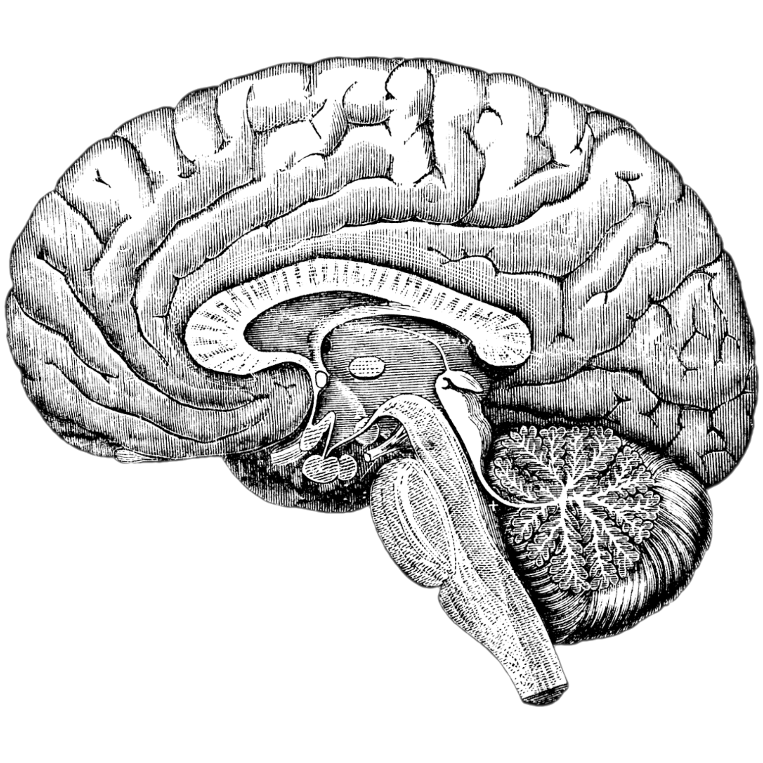

Figure 2: Animal and plant electrical signaling networks. Top Left: Cross section of a human spinal cord, showing regions with different types of signal carrying “cables” (neurons). Top Right: Cross section of a plant root, showing regions with different types of signal carrying “cables”, including phloem, which carries electrical signals, and xylem, which carries hydraulic signals (which can induce electrical signals). Bottom Left: Cross section of a human brain, which is made up of many neural connections. Bottom Right: A tree, which is made up of many phloem (and xylem) connections.
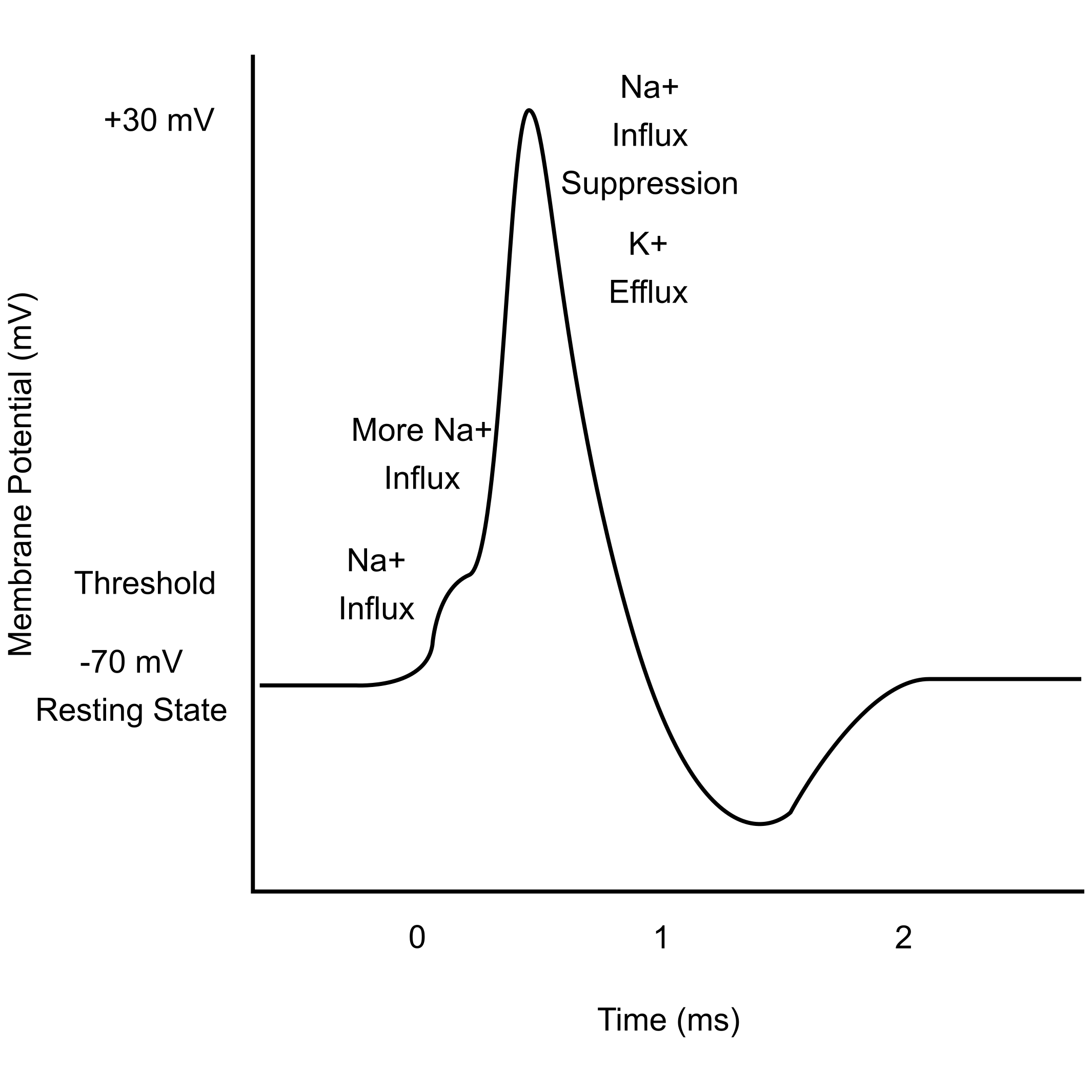
In both plants and animals, information is transmitted through their cable of specialized cells by means of an “action potential” (AP). An action potential is a self-sustaining chain reaction of opening and closing ion channels that occurs along the length of the cable. In animal neurons, the ions involved in this process are K+, Na+, and Ca2+ (Figure 3, Left), while in plant sieve elements the ions involved are K+, Cl-, and Ca2+ [7] (Figure 3, Right). Despite these similarities, with regard to temporal characteristics, plant action potentials are longer in duration than those in animals [8,9] (See time scale, Figure 3).
Animal
Plant
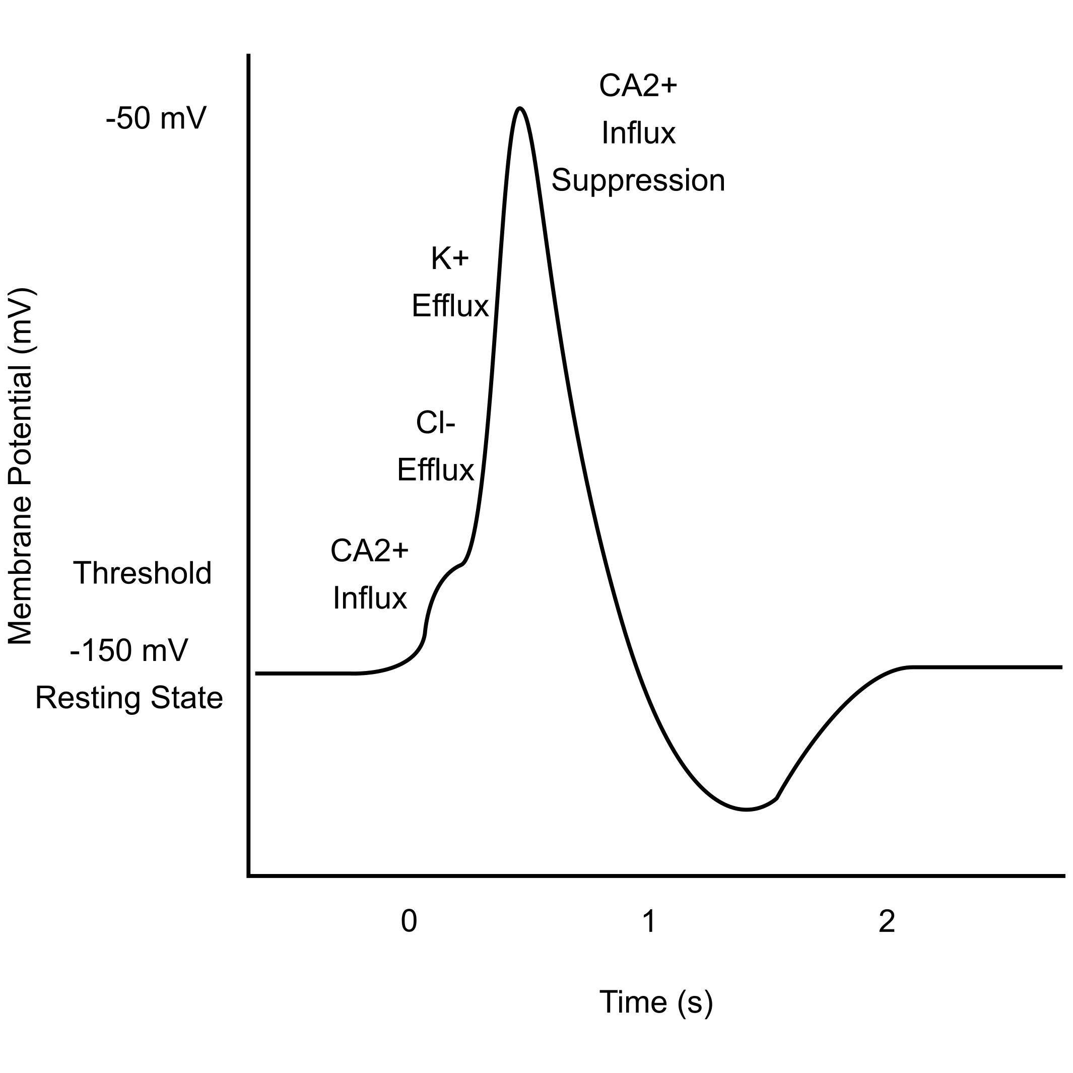
Figure 3: Animal and plant action potentials (APs) [9]
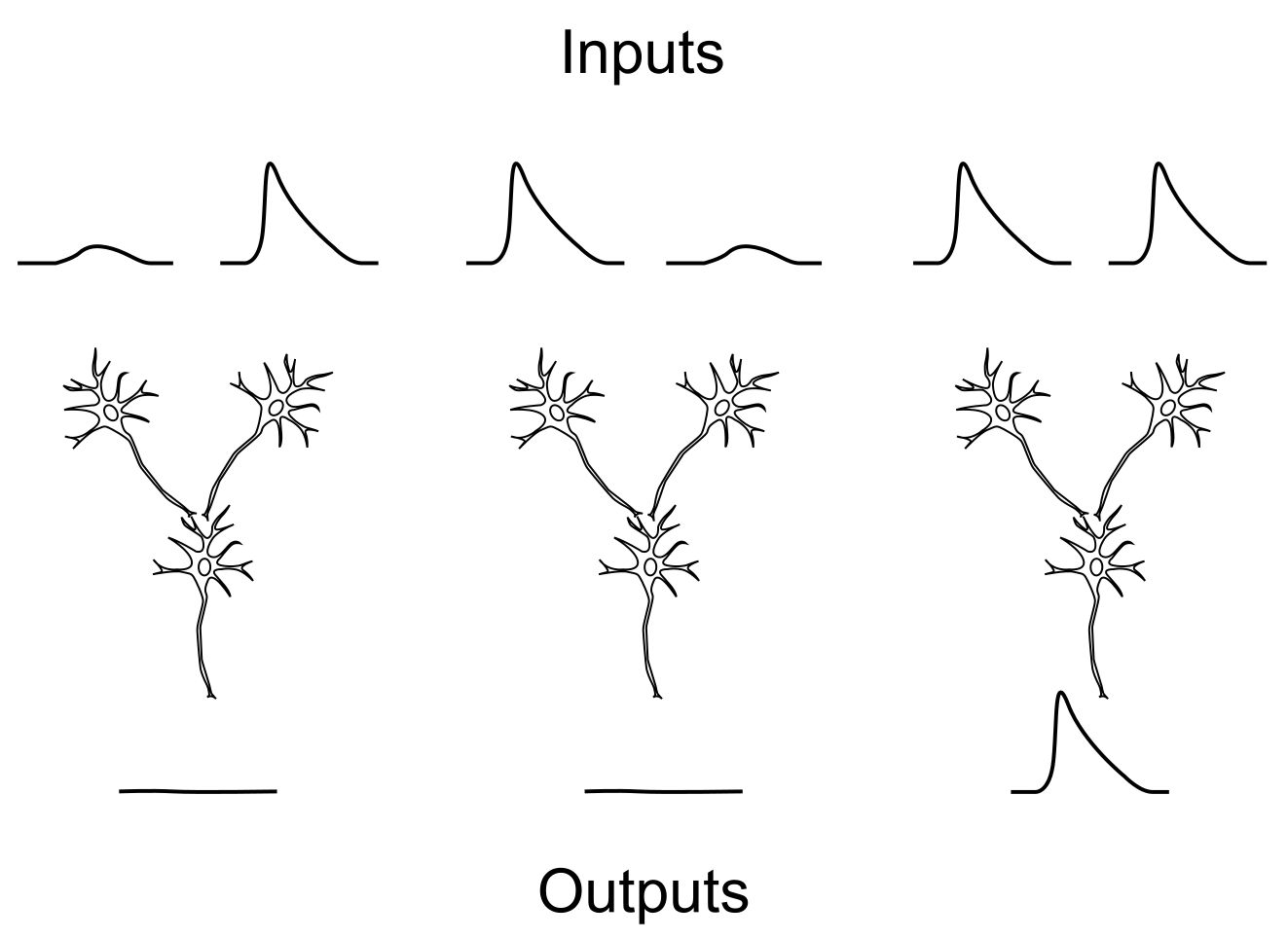
In animals, action potentials are thought to transmit information via temporal and spatial encoding [10]. Temporal encoding means that information is conveyed in the timing, or “firing rate” of APs in a series, and spatial encoding means that information is conveyed in the convergence of APs from multiple distinct sources (Figure 4, Left). Plants have been shown to use both temporal and spatial AP encoding as well. The best known use of temporal encoding in plants is in the venus fly trap [11], however recent studies in more common plants, like sunflowers, have shown signs of this encoding technique as well [12]. Meanwhile, spatial encoding was recently demonstrated at the junction of a plant petiole and stem in mimosa plants (Figure 4, Right) [13].
Animal
Plant
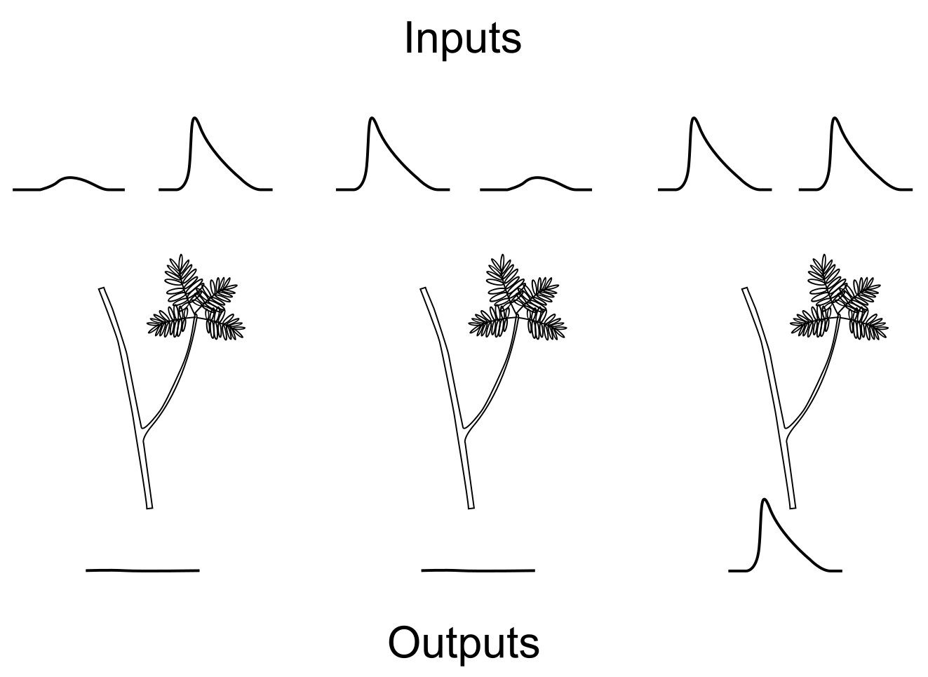
Figure 4: Spatial encoding in animals [10] and plants [13]. Left: A depiction of two “input” neurons (top) sending signals to one “output” neuron (bottom). When both “input” neurons fire, they increase the probability of making the “output” neuron fire. Right: A depiction of a Mimosa pudica plant stem with a side branch. Experiments have shown that the upper stem and side branch act similarly to the top two “input” neurons shown on the left side of the diagram. When stimuli induce electrical signals in both the upper stem and the side branch simultaneously, the probability of the electrical signal propagating into the lower stem below the junction increases.
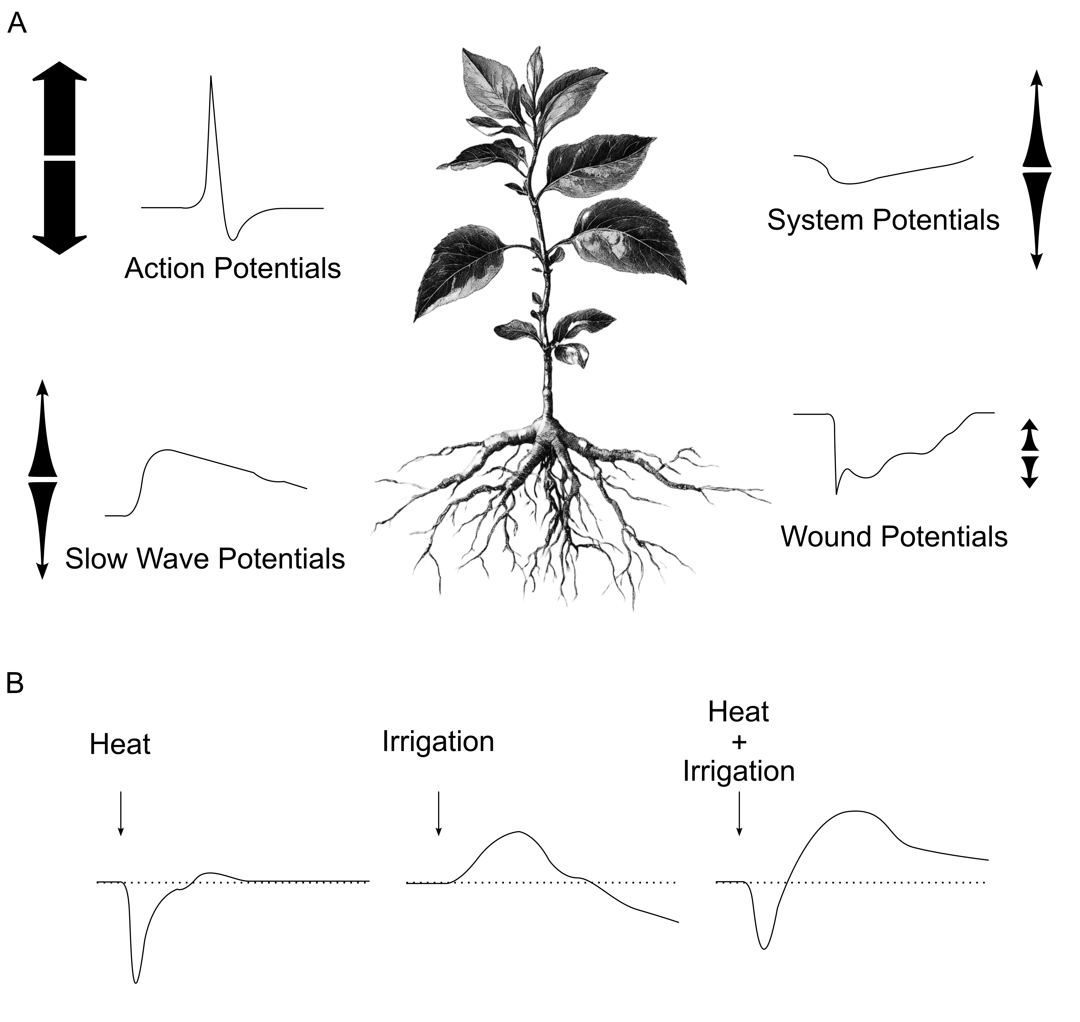
In addition to action potentials, plants also have an assortment of other electrical signals that are combined in various patterns (sometimes along with action potentials) to encode information. These signals, illustrated in Figure 5A, include, “slow wave potentials”, which are caused by hydraulic pressure changes, as well as “system potentials”, and “wound potentials” [14]. These different types of signals can overlap and interact if they occur simultaneously (e.g. due to two simultaneous stimuli), as shown in Figure 5B [15].
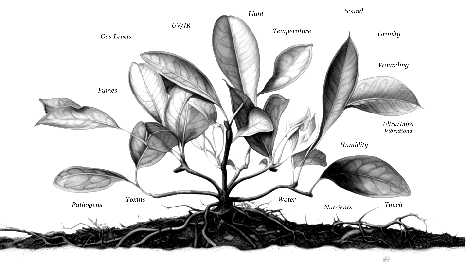
Plants share a significant portion of their internal machinery, and therefore their genome, with humans, and animals in general [6]. As such, many stimuli that negatively affect humans also negatively affect plants. This includes the obvious ones like being bitten or cut, being too hot or too cold, or not having enough water, and it also importantly includes environmental chemical toxins, to which animals and plants share many common sensitivities [16,17]. That’s why researchers have to work hard to find weed killers that don’t also harm humans, and pesticides that don’t also harm plants. Plants are affected by chemical toxins when they bind to microscopic protein receptors, or destroy plant tissue, either of which can then trigger electrical signaling in the plant. As such, plant electrical signals can encode information about chemicals and pathogens in the air, in the soil, and ground water [17–22], in addition to encoding the presence of specific electromagnetic (light) wavelengths [23,24], pollen landing on their sex organs [25,26], touch onset and offset [27,28], wounding [29–31], soil moisture [32], temperature [33,34], and herbivorous insects [35]. This encoded information about various stimuli travels to, and is used by appropriate regions of the plant’s body to regulate various aspects of its metabolism and reproductive processes, including respiration, vascular transport, fertilization, photosynthesis, and even gene expression [7]. Because different parts of the plant body are responsible for each of these senses and functions, signals are constantly being transmitted back and forth between the roots, leaves, and other parts of the plant, creating electrical fluctuations across its body at different times of day, and in different environmental states [12,36,37].
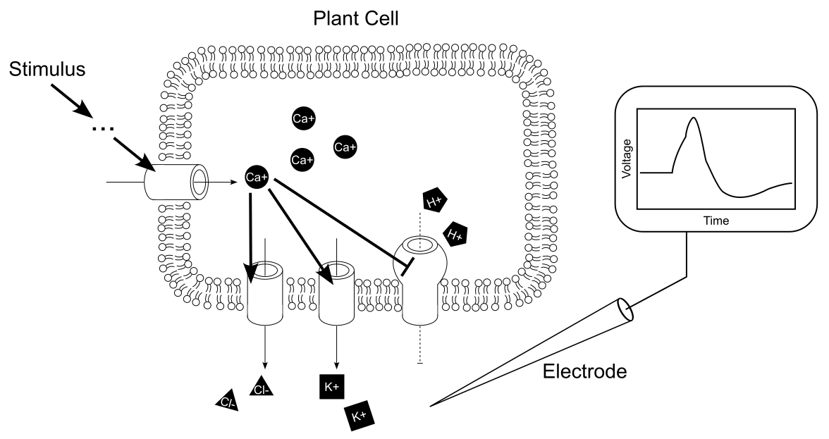
We previously listed stimuli that have been shown in experiments to trigger recognizable responses in plant electrical signals. But what other stimuli in a plant’s environment might also be decodable in this way? The short answer is: any stimulus that induces calcium ion (Ca+) fluctuations inside plant cells has the potential to cause electrical fluctuations that can be detected. These electrical changes could be detected either locally in the cells directly in contact with the stimulus, or, if long distance signal transduction is involved, in regions more distant from the stimulus. This is because Ca+ has been found to be a common element in the translation of most, if not all, stimuli into electrical signals. The way that different stimuli induce this Ca+ fluctuation varies greatly (see “...” in Figure 7), as does the subsequent way in which this induces changes in electrical membrane potentials. A more in depth coverage of this translation of stimuli into electrical signals can be found in several recent papers [38–40].
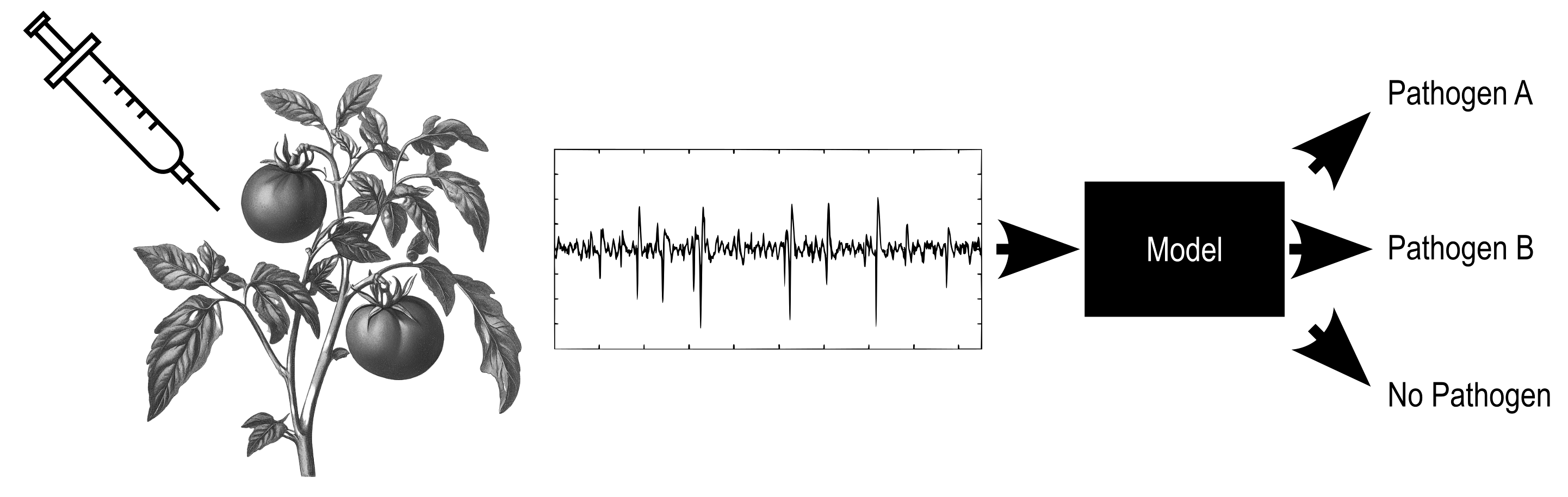
While plant researchers in the past relied on relatively simple analysis of correlations between stimuli and resultant action potentials or variation potentials in electromagnetically shielded laboratory setups, recently researchers have begun using the power of machine learning to allow recording and processing of higher complexity signals in an electrically noisy, real world environment, outside the laboratory. With the use of machine learning, signals recorded from a single electrode connected to a plant stem have been shown to offer insights into whether a plant is 1) water satiated [32,41,42], 2) being preyed on by one or more species of insects [43,44] or fruit eating herbivores [45], 3) deficient in iron [46], nitrogen [47], calcium, or manganese [48], 4) being exposed to liquid toxins [49], or gaseous toxins [49], or 5) even whether a parasitic plant is near a host plant [50]. While these studies showed the ability to detect the presence of individual stimuli, recent studies have also begun showing the ability to distinguish multiple stimuli using a single model. This was first approached by Najdenovska et al. in 2021 [51], and then in subsequent papers evaluating a wide range of stimuli including drought, calcium deficit, and spider mite infestation [51], sodium chloride, sulfuric acid, and ozone [52], wind, heat, red light, and blue light [53], and different pathogens [22]. The accuracy of these multiclass classification models ranged from 73% to >99%, suggesting strong potential for real world application and ongoing extension and refinement of this research (Figure 8).
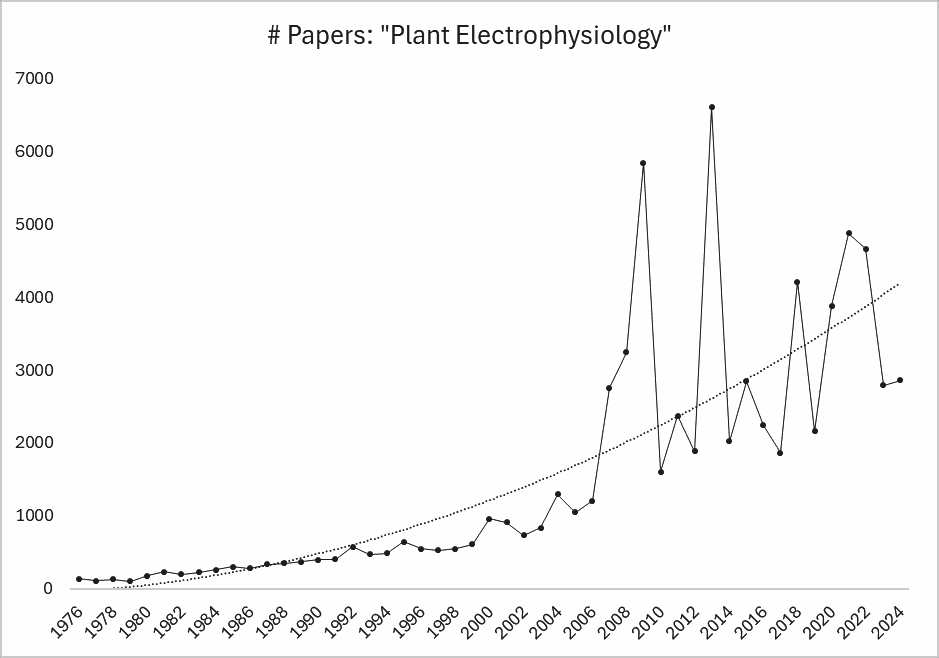
Because of the facilitation of plant electrophysiology research by machine learning techniques, research in the field of plant electrophysiology is rapidly increasing (see Figure 9).
Plants are highly sensitive environmental sensors, simultaneously sampling thousands of features from 1) the soil from which we grow our food, 2) the groundwater from which we drink, and 3) the atmosphere from which we breathe. Furthermore, their non-motile nature also allows plants to establish an intricately intertwined signaling network with a mesh of neighboring plants (and fungi) of the same, and different, species (Figure 9) [54,55]. Finally, remaining in one place also allows plants to be long-term sentinels of specific geographic locations, tracking relative changes over time, and hyperaccumulating environmental toxins that may be present in only trace amounts [56]. We want to tap into this immense, multi-level network of sensors that is distributed across all environmental regimes relevant to human health and productivity.
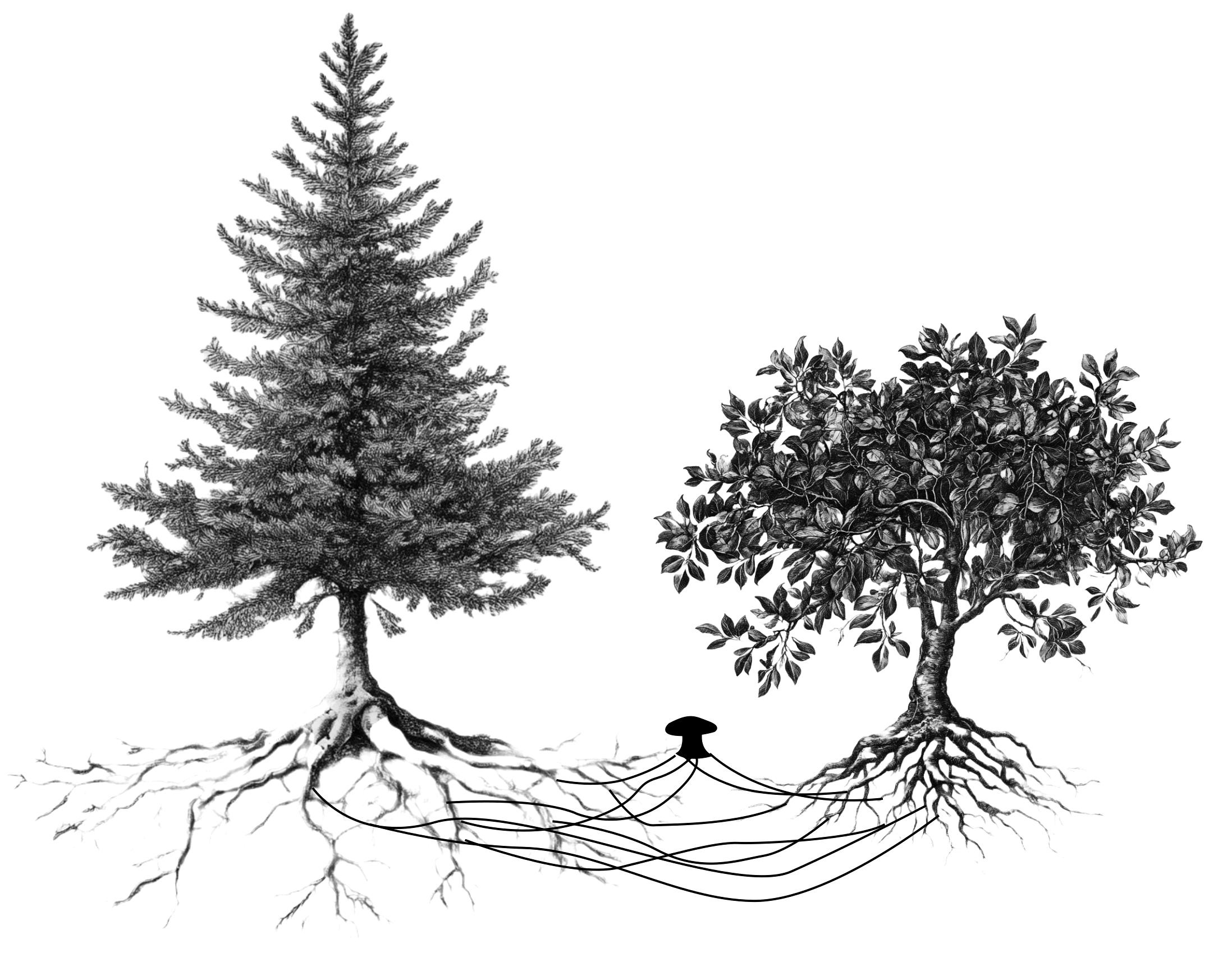
As alluded to previously, plants don’t just relay information within their own bodies, they also share information bidirectionally with neighboring plants via both above ground and subsurface channels. Above ground information transfer is done via electrical signals A) jumping directly across overlapping leaves of neighboring plants [57], and B) possibly triggering the generation of ultrasonic sounds [58] and gaseous chemicals [59–62] that trigger electrical responses in plants within “hearing” / “smelling” range [63]. Meanwhile, subsurface communication between plants involves electrical signals coursing A) through fungal (“mycorrhizal”) networks in the soil, which bridge the gap between neighboring plant roots [55,64,65], B) through the soil itself [66,67], or C) even through naturally grafted / merged (“inosculated”) roots between two plants of the same species [68]. As should be evident from the above, our rapidly increasing understanding of this network of connected plants holds promise for fascinating future discoveries. Indeed, due to their intricate and direct physical connections with neighboring plants (allowed by their non-motile nature), it’s possible that plant-to-plant communication may rival that of many animals, allowing a valuable synthesis of information from an “internet of plants”.
We’ve shown that plants are highly sensitive to their surroundings, acting as sophisticated biological multi-sensors, with an ability to electrically process and communicate that information, both internally and with neighboring plants. As scientific research continues and accelerates, it seems that we're just getting started in understanding the full exciting extent of plant capabilities.
1. Lemieux, C., Otis, C. & Turmel, M. Ancestral chloroplast genome in Mesostigma viride reveals an early branch of green plant evolution. Nature 403, 649–652 (2000).
2. Leliaert, F. et al. Phylogeny and Molecular Evolution of the Green Algae. Crit. Rev. Plant Sci. 31, 1–46 (2012).
3. Kandasamy, M. K., McKinney, E. C., Roy, E. & Meagher, R. B. Plant Vegetative and Animal Cytoplasmic Actins Share Functional Competence for Spatial Development with Protists[W][OA]. Plant Cell 24, 2041–2057 (2012).
4. Haraguchi, T., Duan, Z., Tamanaha, M., Ito, K. & Tominaga, M. Diversity of Plant Actin–Myosin Systems. in The Cytoskeleton: Diverse Roles in a Plant’s Life (eds. Sahi, V. P. & Baluška, F.) 49–61 (Springer International Publishing, Cham, 2019). doi:10.1007/978-3-030-33528-1_4.
5. Renzaglia, K., Villarreal A., J. & Garbary, D. Morphology supports the setaphyte hypothesis: mosses plus liverworts form a natural group. Bryophyte Divers. Evol. 40, 11–017 (2018).
6. Do People and Bananas Really Share 50 Percent of the Same DNA? HowStuffWorks https://science.howstuffworks.com/life/genetic/people-bananas-share-dna.htm (2021).
7. Plant Electrophysiology: Signaling and Responses. (Springer Berlin Heidelberg, Berlin, Heidelberg, 2012). doi:10.1007/978-3-642-29110-4.
8. Brownlee, C. Plant physiology: Anatomy of a plant action potential. Curr. Biol. 32, R1000–R1002 (2022).
9. Lee, J. & Calvo, P. The potential of plant action potentials. Synthese 202, 176 (2023).
10. Auge, D., Hille, J., Mueller, E. & Knoll, A. A Survey of Encoding Techniques for Signal Processing in Spiking Neural Networks. Neural Process. Lett. 53, (2021).
11. Jacobson, S. L. Receptor Response in Venus’s Fly-Trap. J. Gen. Physiol. 49, 117–129 (1965).
12. Zawadzki, T., Dziubiśka, H. & Davies, E. Characteristics of action potentials generated spontaneously in Helianthus annuus. Physiol. Plant. 93, 291–297 (1995).
13. de Bakker, J. M. T. & Coronel, R. Summation of activation at the branch-stem transition of Mimosa pudica; a comparison with summation in cardiac tissue. PLOS ONE 18, e0286103 (2023).
14. Huber, A. E. & Bauerle, T. L. Long-distance plant signaling pathways in response to multiple stressors: the gap in knowledge. J. Exp. Bot. 67, 2063–2079 (2016).
15. Vuralhan-Eckert, J., Lautner, S. & Fromm, J. Effect of simultaneously induced environmental stimuli on electrical signalling and gas exchange in maize plants. J. Plant Physiol. 223, 32–36 (2018).
16. LaLone, C. A. et al. International Consortium to Advance Cross-Species Extrapolation of the Effects of Chemicals in Regulatory Toxicology. Environ. Toxicol. Chem. 40, 3226–3233 (2021).
17. Volkov, A. G., Collins, D. J. & Mwesigwa, J. Plant electrophysiology: pentachlorophenol induces fast action potentials in soybean. Plant Sci. 153, 185–190 (2000).
18. Shvetsova, T. et al. Soybean electrophysiology: effects of acid rain. Plant Sci. 162, 723–731 (2002).
19. Mwesigwa, J., Collins, D. J. & Volkov, A. G. Electrochemical signaling in green plants: effects of 2,4-dinitrophenol on variation and action potentials in soybean. Bioelectrochemistry 51, 201–205 (2000).
20. Bricchi, I., Bertea, C. M., Occhipinti, A., Paponov, I. A. & Maffei, M. E. Dynamics of Membrane Potential Variation and Gene Expression Induced by Spodoptera littoralis, Myzus persicae, and Pseudomonas syringae in Arabidopsis. PLOS ONE 7, e46673 (2012).
21. Simmi, F. Z., Dallagnol, L. J., Ferreira, A. S., Pereira, D. R. & Souza, G. M. Electrome alterations in a plant-pathogen system: Toward early diagnosis. Bioelectrochemistry 133, 107493 (2020).
22. Zaiosc Simmi, F., José Dallagnol, L., Oliveira Almeida, R., da Rosa Dorneles, K. & Maia Souza, G. Barley systemic bioelectrical changes detect pathogenic infection days before the first disease symptoms. Comput. Electron. Agric. 209, 107832 (2023).
23. Nishizaki, Y. Blue Light Pulse-Induced Transient Changes of Electric Potential and Turgor Pressure in the Motor Cells of Phaseolus vulgaris L. Plant Cell Physiol. 29, 1041–1046 (1988).
24. Volkov, A. G. et al. Bioelectrochemical signaling in green plants induced by photosensory systems. Bioelectrochemistry 63, 91–94 (2004).
25. Sinyukhin, A. M. & Britikov, E. A. Action Potentials in the Reproductive System of Plants. Nature 215, 1278–1280 (1967).
26. Fromm, J., Hajirezaei, M. & Wilke, I. The Biochemical Response of Electrical Signaling in the Reproductive System of Hibiscus Plants. Plant Physiol. 109, 375–384 (1995).
27. Agosti, R. D. Touch-induced action potentials in Arabidopsis thaliana. Arch. Sci. (2014).
28. Howell, A. H. et al. Pavement cells distinguish touch from letting go. Nat. Plants 1–6 (2023) doi:10.1038/s41477-023-01418-9.
29. Davies, E. Action potentials as multifunctional signals in plants: a unifying hypothesis to explain apparently disparate wound responses. Plant Cell Environ. 10, 623–631 (1987).
30. Rhodes, J. D., Thain, J. F. & Wildon, D. C. Signals and Signalling Pathways in Plant Wound Responses. in Communication in Plants: Neuronal Aspects of Plant Life (eds. Baluška, F., Mancuso, S. & Volkmann, D.) 391–401 (Springer, Berlin, Heidelberg, 2006). doi:10.1007/978-3-540-28516-8_26.
31. Roblin, G. Analysis of the Variation Potential Induced by Wounding in Plants. Plant Cell Physiol. 26, 455–461 (1985).
32. Ríos-Rojas, L., Tapia, F. & Gurovich, L. A. Electrophysiological assessment of water stress in fruit-bearing woody plants. J. Plant Physiol. 171, 799–806 (2014).
33. Sibaoka, T. Transmission of action potentials in Biophytum. Bot. Mag. Shokubutsu-Gaku-Zasshi 86, 51–61 (1973).
34. Sambeek, J. W. V. & Pickard, B. G. Mediation of rapid electrical, metabolic, transpirational, and photosynthetic changes by factors released from wounds. I. Variation potentials and putative action potentials in intact plants. Can. J. Bot. 54, 2642–2650 (1976).
35. Volkov, A. G. & Haack, R. A. Insect-induced biolectrochemical signals in potato plants. Bioelectrochem. Bioenerg. 37, 55–60 (1995).
36. Stolarz, M. & Dziubińska, H. Spontaneous action potentials and circumnutation in Helianthus annuus. Acta Physiol. Plant. 39, 234 (2017).
37. Stolarz, M. & Trębacz, K. Spontaneous rapid leaf movements and action potentials in Mimosa pudica. Physiol. Plant. 173, 1882–1888 (2021).
38. Sanders, D., Pelloux, J., Brownlee, C. & Harper, J. F. Calcium at the Crossroads of Signaling. Plant Cell 14, s401–s417 (2002).
39. Choi, W.-G., Hilleary, R., Swanson, S. J., Kim, S.-H. & Gilroy, S. Rapid, Long-Distance Electrical and Calcium Signaling in Plants. Annu. Rev. Plant Biol. 67, 287–307 (2016).
40. Mudrilov, M., Ladeynova, M., Grinberg, M., Balalaeva, I. & Vodeneev, V. Electrical Signaling of Plants under Abiotic Stressors: Transmission of Stimulus-Specific Information. Int. J. Mol. Sci. 22, 10715 (2021).
41. Yu, R., Wu, Y. & Xing, D. Can Electrophysiological Parameters Substitute for Growth, and Photosynthetic Parameters to Characterize the Response of Mulberry and Paper Mulberry to Drought? Plants 10, 1772 (2021).
42. Tran, D. et al. Electrophysiological assessment of plant status outside a Faraday cage using supervised machine learning. Sci. Rep. 9, 17073 (2019).
43. Vivent. Optimizing crop production using real-time plant electrophysiology. https://www.youtube.com/watch?v=45Au5NAJqrQ (2023).
44. Najdenovska, E. et al. Classification of Plant Electrophysiology Signals for Detection of Spider Mites Infestation in Tomatoes. Appl. Sci. 11, 1414 (2021).
45. Reissig, G. N. et al. Fruit Herbivory Alters Plant Electrome: Evidence for Fruit-Shoot Long-Distance Electrical Signaling in Tomato Plants. Front. Sustain. Food Syst. 5, (2021).
46. Tran, D. & Camps, C. Early Diagnosis of Iron Deficiency in Commercial Tomato Crop Using Electrical Signals. Front. Sustain. Food Syst. 5, (2021).
47. González I Juclà, D., Najdenovska, E., Dutoit, F. & Raileanu, L. E. Detecting stress caused by nitrogen deficit using deep learning techniques applied on plant electrophysiological data. Sci. Rep. 13, 9633 (2023).
48. Sai, K., Sood, N. & Saini, I. Classification of various nutrient deficiencies in tomato plants through electrophysiological signal decomposition and sample space reduction. Plant Physiol. Biochem. 186, 266–278 (2022).
49. Chatterjee, S. K. et al. Exploring strategies for classification of external stimuli using statistical features of the plant electrical response. J. R. Soc. Interface 12, 20141225 (2015).
50. Parise, A. G. et al. Detection of Different Hosts From a Distance Alters the Behaviour and Bioelectrical Activity of Cuscuta racemosa. Front. Plant Sci. 12, (2021).
51. Najdenovska, E. et al. Identifying General Stress in Commercial Tomatoes Based on Machine Learning Applied to Plant Electrophysiology. Appl. Sci. 11, 5640 (2021).
52. Bhadra, N., Chatterjee, S. K. & Das, S. Multiclass classification of environmental chemical stimuli from unbalanced plant electrophysiological data. PLOS ONE 18, e0285321 (2023).
53. Buss, E. et al. Stimulus classification with electrical potential and impedance of living plants: comparing discriminant analysis and deep-learning methods. Bioinspir. Biomim. 18, 025003 (2023).
54. Geara, H., Valdur, K.-A. & Must, I. Mycelium Bridge as a Living Electrical Conductor: Access Point to Soil Infosphere. in Biomimetic and Biohybrid Systems (eds. Meder, F., Hunt, A., Margheri, L., Mura, A. & Mazzolai, B.) 335–347 (Springer Nature Switzerland, Cham, 2023). doi:10.1007/978-3-031-39504-8_23.
55. Thomas, M. A. & Cooper, R. L. Building bridges: mycelium–mediated plant–plant electrophysiological communication. Plant Signal. Behav. 17, 2129291 (2022).
56. Behmer, S. T. et al. Metal hyperaccumulation in plants: mechanisms of defence against insect herbivores. Funct. Ecol. 19, 55–66 (2005).
57. Szechyńska-Hebda, M. et al. Aboveground plant-to-plant electrical signaling mediates network acquired acclimation. Plant Cell 34, 3047–3065 (2022).
58. Mishra, R. C., Ghosh, R. & Bae, H. Plant acoustics: in the search of a sound mechanism for sound signaling in plants. J. Exp. Bot. 67, 4483–4494 (2016).
59. Baldwin, I. T. & Schultz, J. C. Rapid changes in tree leaf chemistry induced by damage: evidence for communication between plants. Science 221, 277–279 (1983).
60. Maffei, M. E., Gertsch, J. & Appendino, G. Plant volatiles: production, function and pharmacology. Nat. Prod. Rep. 28, 1359–1380 (2011).
61. Sugimoto, K., Matsui, K. & Takabayashi, J. Uptake and conversion of volatile compounds in plant–plant communication. Deciphering Chem. Lang. Plant Commun. 305–316 (2016).
62. Zebelo, S. A., Matsui, K., Ozawa, R. & Maffei, M. E. Plasma membrane potential depolarization and cytosolic calcium flux are early events involved in tomato (Solanum lycopersicon) plant-to-plant communication. Plant Sci. 196, 93–100 (2012).
63. Loreto, F. & D’Auria, S. How do plants sense volatiles sent by other plants? Trends Plant Sci. 27, 29–38 (2022).
64. Song, Y. Y. et al. Interplant communication of tomato plants through underground common mycorrhizal networks. PloS One 5, e13324 (2010).
65. Babikova, Z., Johnson, D., Bruce, T., Pickett, J. & Gilbert, L. How rapid is aphid-induced signal transfer between plants via common mycelial networks? Commun. Integr. Biol. 6, e25904 (2013).
66. Volkov, A. G. & Shtessel, Y. B. Electrical signal propagation within and between tomato plants. Bioelectrochemistry 124, 195–205 (2018).
67. G Volkov, A. & B Shtessel, Y. Electrotonic signal transduction between Aloe vera plants using underground pathways in soil: Experimental and analytical study. AIMS Biophys. 4, (2017).
68. Mylo, M. D. et al. Conjoining Trees for the Provision of Living Architecture in Future Cities: A Long-Term Inosculation Study. Plants 12, 1385 (2023).
